1. Distribution- face, neck and sun-exposed areas
of the upper trunk and arms
2. Sunlight sensitivity
Clinical Presentation
Lupus erythematosus (LE) is a disease that has a very broad
spectrum of clinical symptoms and signs. The spectrum is
continuous, but it is convenient to consider four points on the
spectrum as if they were four separate conditions.
Only the discoid-type skin lesions possess all of the
characteristics of papulosquamous disease. The skin lesions that
occur in patients with systemic disease lack one or more
papulosquamous characteristics and thus overlap morphologically
with diseases of the vascular reaction and eczematous disease
groups.
The lesions in all types of LE are, in general, correlated with
sunlight exposure. They are primarily found on the sun-exposed
portions of the body, and in many cases the patient will have
noted the development of new lesions (or the worsening of old
lesions) following one or more episodes of sunlight exposure.
Nevertheless, a specific history of photosensitivity is often
lacking.
The patches and plaques of LE are generally asymptomatic, but a
sensation of swelling and burning is sometimes described by
patients with the lesions of systemic LE.
The diagnosis of LE involving the skin is often possible on the
basis of clinical examination. Confirmation should be obtained
through biopsy, however. Biopsy of skin lesions for direct
immunofluorescence (the lupus band test) is particularly helpful,
since it regularly reveals the deposition of complement and
immunoglobulin at the dermal-epidermal junction. Similar deposits
can also be identified in the nonlesional skin in about 70% or 80%
of those patients who have systemic disease. Serologic tests for
fluorescent antinuclear antibodies (FANA) are also useful in the
diagnosis of LE.
Discoid Lupus Erythematosus
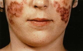
The skin lesions of discoid LE consist of sharply marginated,
erythematous plaques 1 to 4 cm in diameter. Large lesions are
often annular with a thin erythematous scaling border surrounding
a white, scarred center. Smaller lesions are solid (as opposed to
annular) that-topped papules and plaques diffusely covered with
scale. lesions of discoid LE occur anywhere on the face but are
most often found on the lateral cheeks, particularly at the
Lawline. The distribution, although usually bilateral, is often
not symmetrical. Lesions in the scalp occur as sharply localized
patches of hair loss . Gray-white plaques are sometimes found on
the lips and oral mucous membranes. Discoid LE occurs with
approximately equal frequency in men and women. The disease
develops at any point from childhood to late adult life . Systemic
symptoms and signs are almost always absent in patients with
discoid lesions. The incidence in men and women is almost equal.
Antinuclear antibodies as determined by fluorescent antinuclear
antibody (FANA) tests are usually negative.
Disseminated Discoid Lupus Erythematosus
There is, however, less tendency for central clearing and for
scarring. Moreover, the distribution pattern is more extensive;
lesions are found on the sun-exposed surfaces of the arms and
hands as well as on the face. Scarring alopecia is not often
present. Ten percent to 20% of patients with this type of LE will
have a positive F ANA test.
Subacute Lupus Erythematosus
Often, the term subacute cutaneous is used for this type of
disease. Two types of lesions may occur. The first consists of
lesions that are annular plaques 2 to 10 cm in diameter. The ring
of the annulus is 3 to 5 mIll wide and has little or no scale. The
central portion appears as normal skin. Coalescence of these
lesions to form larger plaques with a gyrate configuration
sometimes occurs . The second type consists of solid plaques that
resemble psoriasis. Differentiation from psoriasis is possible
because the margination may be a bit less distinct, the scale size
is smaller, and there is no evidence of the Koebner phenomenon.
Both types of subacute LE are distributed primarily on the upper
trunk and lateral arms; the face is usually spared. Patients with
subacute LE usually have a positive FANA test; more specifically,
Ro (SSA) antibodies are regularly present.
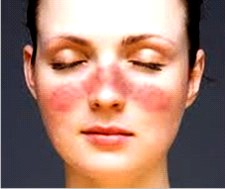
Acute Systemic Lupus Erythematosus
The cutaneous hallmark of acute systemic LE is the presence of
symmetrical, poorly marginated, erythematous plaques on the upper
malar prominences. The bridge of the nose may also be involved.
Scale formation in this so-called butterfly eruption is minimal,
and the plaque is usually somewhat edematous. When lesions occur
other than on the face, there is a marked tendency for
coalescence, as opposed to the smaller discrete lesions of discoid
LE. Hair loss, when it occurs, is diffuse rather than localized.
Mucous membrane lesions occur in about 25% of patients, they are
identical with those seen in discoid LE .
Both sides of the hands are regularly involved. Small patches of
erythema are located on the dorsal surface of the phalanges, but
the area over the knuckles is spared. Reddening and telangiectasia
are frequently present in a narrow band at the posterior nail
folds. The palmar surfaces of the hands are often violaceous. This
color change is particularly notable over the tips of the fingers
and on the thenar and hypothenar eminences. Small, bright red,
blanching macules or pinpoint violaceous vasculitic lesions may be
superimposed against these duskier color changes. Women with this
condition outnumber men with this condition by a considerable
margin.
Course and Prognosis
The course and prognosis of LE correlate rather well with various
types of cutaneous lesions. Patients with discoid lesions confined
to the face may have a few minor laboratory abnormalities but
rarely, if ever, have symptoms and signs of systemic disease.
Moreover, 95% of such patients will have a normal life span with
only cutaneous morbidity as a manifestation of their disease.
Patients with disseminated discoid skin lesions usually have a
number of minor laboratory abnormalities, but they, too, rarely
develop significant systemic disease.
Patients with lesions of subacute LE often have fever and
arthralgia, but cardiac, central nervous system, and renal
involvement are usually mild or absent. Patients with lesions of
acute LE are highly likely to have serious systemic symptoms and
signs.
The lesions of discoid LE heal with scarring and sometimes “burn
out” altogether after 10 to 20 years of activity. The lesions of
subacute and acute LE heal without scarring. These latter lesions
tend to mirror the activity of the underlying systemic disease;
i.e., they fade during periods of remission and reappear during
exacerbations.
Pathogenesis
The cause of skin lesions in LE is not known. In most instances,
sunlight seems to play an important precipitating role. Exposure
to the 280- to 320-nm wavelengths
of ultraviolet light (the UVB, or sunburn, spectrum) presumably
leads to DNA damage in epithelial cells. It is hypothesized that
this modified DNA then acts as a new antigen that stimulates the
production of “autoimmune” antibodies. The production of these
antibodies by B cells may be enhanced by a reduction in suppressor
T cells. Antibodies, once formed, are deposited along with
complement in the skin and other organs. Genetic factors, as
manifest by the frequency of familial cases and the presence of
certain HLA patterns, are undoubtedly important. Since the more
serious forms of LE occur primarily in women, it is suspected that
the presence of estrogens (or absence of androgens) may playa
role.
Therapy
All patients should be protected from ultraviolet light
irradiation in the UVB (sunburn) spectrum. This can be
accomplished rather well by regular application of sunscreens with
a high sun protective factor (SPF) . In addition, protective
clothing and a change in lifestyle that moves outdoor activities
to the beginning and end of the day are recommended.
The cutaneous lesions of LE resolve with steroid treatment.
Discoid lesions respond inconsistently to topical steroids but do
improve with intralesional injections of triamcinolone. The
lesions of subacute and acute LE clear if associated systemic
disease is treated with systemic steroids. The oral administration
of hydroxychloroquine (Plaquenil) in a daily dose of 200 to 400 mg
is very helpful in the treatment of all types of lesions
Growing Stronger, Growing
Better
Healthcare Provider
Lupus erythematosus - treatment of Lupus
erythematosus, Lupus erythematosus types, Disease medicines, Lupus
erythematosus symptoms, Lupus erythematosus and Disease symptoms, Lupus
erythematosus symptoms Disease and diagnosis, Symptoms and Solutions, Signs
and Symptoms, type of Lupus erythematosus, cause common, common Lupus
erythematosus, Lupus erythematosus List, causes list, Infectious Lupus
erythematosus, Causes, Diseases , Types, Prevention, Treatment and Facts,
Lupus erythematosus information, Lupus erythematosus: Definition, Lupus
erythematosus names, medical Lupus erythematosus, medical Lupus
erythematosus and disorders, cell Lupus erythematosus, Lupus erythematosus
Worldwide, Lupus erythematosus Research, Lupus erythematosus Control, Lupus
erythematosus Center, Digestive Lupus erythematosus Week, Information about
Lupus erythematosus, causes of different Lupus erythematosus, Lupus
erythematosus Articles, Lupus erythematosus and conditions, Health and Lupus
erythematosus, Lupus erythematosus Patients, Lupus erythematosus and
Sciences, causes of alzheimer's Lupus erythematosus, Lupus erythematosus
causes, alternative medicine heart Lupus erythematosus, body ailments, Lupus
erythematosus medicines, medical antiques, type of blood Lupus erythematosus


 hs
of ultraviolet light (the UVB, or sunburn, spectrum) presumably
leads to DNA damage in epithelial cells. It is hypothesized that
this modified DNA then acts as a new antigen that stimulates the
production of “autoimmune” antibodies. The production of these
antibodies by B cells may be enhanced by a reduction in suppressor
T cells. Antibodies, once formed, are deposited along with
complement in the skin and other organs. Genetic factors, as
manifest by the frequency of familial cases and the presence of
certain HLA patterns, are undoubtedly important. Since the more
serious forms of LE occur primarily in women, it is suspected that
the presence of estrogens (or absence of androgens) may playa
role.
hs
of ultraviolet light (the UVB, or sunburn, spectrum) presumably
leads to DNA damage in epithelial cells. It is hypothesized that
this modified DNA then acts as a new antigen that stimulates the
production of “autoimmune” antibodies. The production of these
antibodies by B cells may be enhanced by a reduction in suppressor
T cells. Antibodies, once formed, are deposited along with
complement in the skin and other organs. Genetic factors, as
manifest by the frequency of familial cases and the presence of
certain HLA patterns, are undoubtedly important. Since the more
serious forms of LE occur primarily in women, it is suspected that
the presence of estrogens (or absence of androgens) may playa
role.